3D-Printed Tools for Biomechanics Research
During my time in the Musculoskeletal Biomechanics Laboratory (MBL), I have designed several 3D-printed tools to assist with laboratory tasks. Find them below.
Cartilage Separator
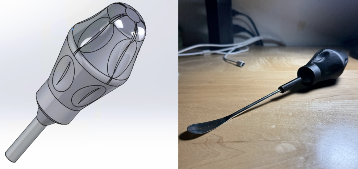
This custom cartilage separation tool is a handheld instrument designed to enable controlled delamination of articular cartilage from underlying bone. I designed and 3D printed a handle to hold a modified screwdriver end. Its rounded filleted working edge conforms to the curved bone–cartilage interface, redistributing contact and reducing localized stress compared to the prior flat, angular tools. This shape minimizes the force required for insertion, improves separation uniformity, and lowers the risk of surface gouging or tissue damage. The contoured handle enhances grip during sample preparation, making the tool better suited for consistent mechanical testing workflows than improvised alternatives such as flathead screwdrivers.
Caliper Alignment Jig
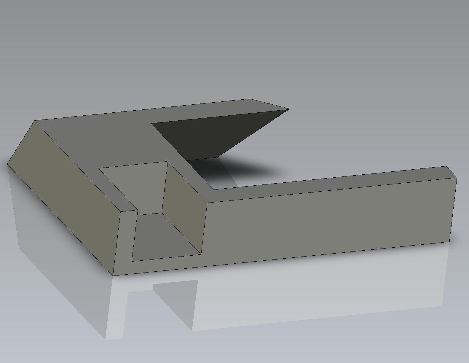
This custom caliper stabilization fixture is a purpose-built tool designed to hold a mechanical caliper steady during microtome-based sample trimming and measurement. I designed and 3D printed the fixture to conform closely to the microtome’s geometry, providing a snug, repeatable interface for caliper placement. Its flat reference surface maintains level alignment and constrains unwanted motion, reducing angular misalignment and measurement variability compared to freehand positioning. By stabilizing the measurement process, the fixture is better suited for consistent dimensional assessment in mechanical testing workflows than ad hoc supports or manual alignment.
Cartilage Sample Holder
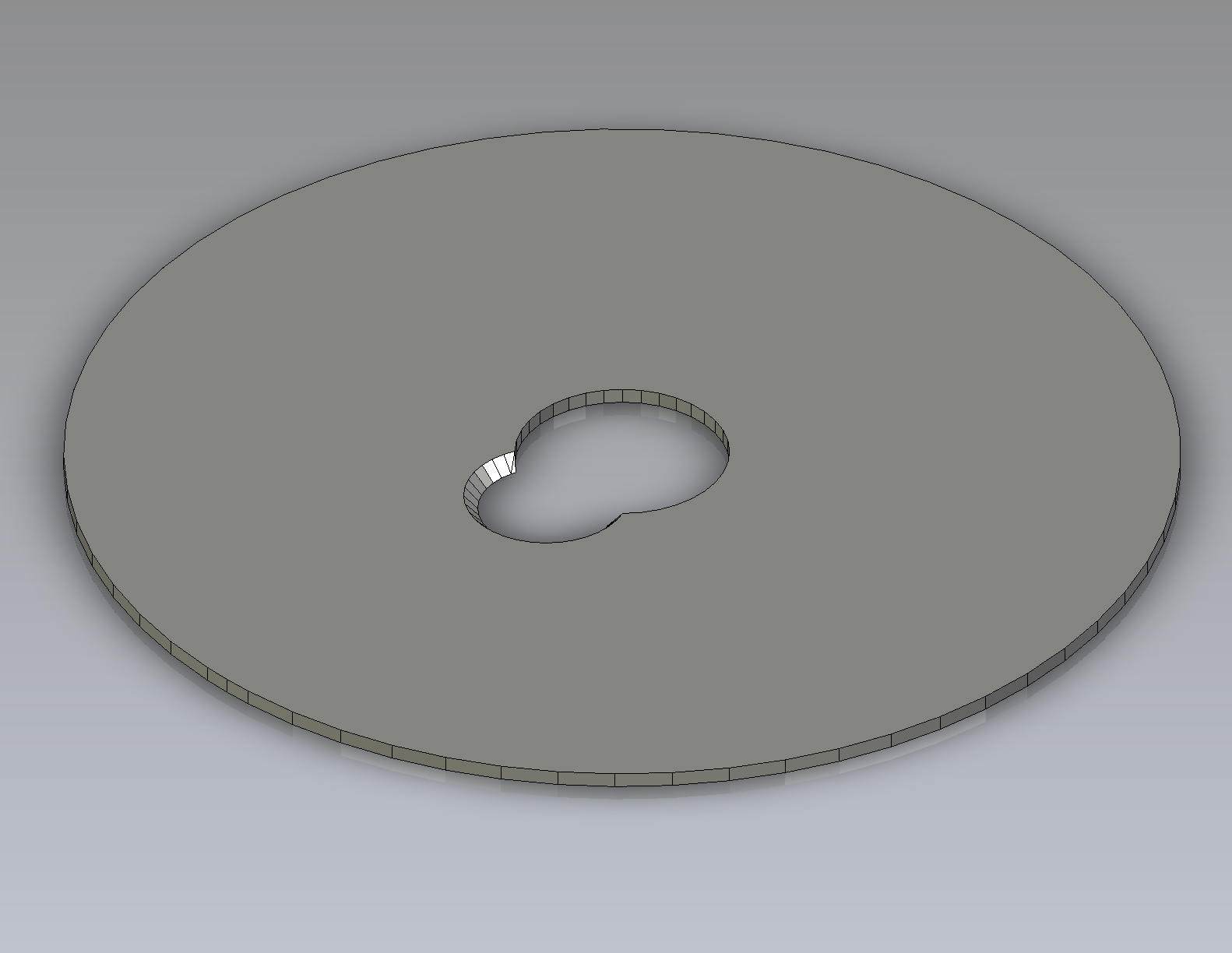
This custom cartilage plug holder is a PLA printed fixture designed to provide repeatable lateral constraint during indentation (confined compression) testing of 10 mm diameter cartilage samples. I designed and 3D printed the holder to interface directly with the existing steel test stage, eliminating the need to glue individual samples and thereby reducing handling time and avoiding potential chemical interaction with the tissue’s superficial zone. All internal and external edges were filleted to prevent sharp stress concentrations or uneven compression at the sample boundary.